multiple tiny echogenic foci in spleen
- kathy garver clearcaptions commercial
- December 11, 2022
b.) Case reports of two patients, Clinical case report: Sclerosing hemangioma of the liver, a rare but great mimicker, Imaging of acute pancreatitis and its complications. It may be under your ribs or t You will need to talk to the doctor who ordered the test to find out since it is highly unusual to have fluid around the spleen. b.) ultrasound of liver, spleen, and pancreas are ok. We conducted a retrospective analysis of all children who had liver and/or spleen abscesses on abdominal ultrasonography admitted to Bintulu Hospital in Sarawak, Malaysia, from January 2014 until December 2018. Would you like email updates of new search results? official website and that any information you provide is encrypted After thoroughly evaluating the LUQ, only a fraction of splenic tissue can be identified. 2023 Dotdash Media, Inc. All rights reserved. a.) 2 . In our experience, infectious and inflammatory diseases account for most cases of multifocal splenic lesions. Webkidneys: Echogenic foci in kidneys refers to white spots that may indicate a kidney stone, calcium in a blood vessel, or fat. m. tumba de Cristbal Coln 266-270, Radiology Case Reports, Volume 11, Issue 2, 2016, pp. 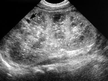 Bezerra AS, D'ippolito G, Caldana RP et-al. a.) Roubidoux MA. defense against disease In this review, the typical splenic abnormalities that can be diagnosed with imaging with a high degree of confidence are illustrated. Nihon Igaku Hoshasen Gakkai Zasshi. This is a common finding of no importance. Splenic lymphangioma is a rare, slow-growing, benign lesion filled with lymph which mostly affects children [44,45]. No treatment is required for this condition.
  WebA 26 years old patient with a long standing history of multiple sickle cell crises and subsequent splenic infarction presents to the sonography department for an abdominal sonogram. Siderotic nodules in the spleen: MR imaging of portal hypertension. Imaging patterns in non-traumatic spleen lesions in adults-a review. Register; Login; EN . splenic hematoma 9. A second patient with mucinous appendiceal neoplasm with peritoneal metastases was studied. HIV bloodwork is perfect. Vascular neoplasms of the spleen represent the majority of the nonhematologic/nonlymphoid neoplasms and commonly produce multifocal lesions. To learn more, please visit our. . Advertisement . Tomato Flu: Symptoms, Causes And Everything We Know So Far, Mother's Day 2022: Mothers - A Boon From God, Countries In WHO South-East Asia Region Renew Commitment To Eliminate Malaria By 2030, Elimination Of Lymphatic Filariasis: Here's How Karnataka Health Officials Are Ensuring Lymphatic Filariasis Doesn't Spread, Urgently Address Gaps In Cancer Care: WHO. Part 2: Complications of acute pancreatitis. 2-11). Bookshelf Melioidosis was confirmed by culture in 9 (17%) children; small occult splenic abscesses were present in all cases. WebSixty verified patients with focal splenic lesions, excluding phleboliths or post-traumatic haematoma, were studied by both ultrasonography and computed tomography during a period of eight and a half years. b.) d.) the spleen is consisted the largest lymphatic organ, c.) the spleen has a convex inferior margin and a concave superior border, The splenic vein joins with what structure posterior to the pancreatic neck to the form the portal vein? Had an ultrasound done last week and the results showed multiple echogenic foci identified in the liver and spleen. Heren, we report a case of splenic IMT with histological correlation. Medicine (Baltimore). WebMultiple, small echogenic foci scattered throughout the spleen in a patient with a history of toxoplasmosis most likely represents: a.) d.) lateral aspect of the pancreatic body and tail, a.) In approximately 1 out of every 20 to 30 pregnancies, an echogenic focus or foci is discovered in a second-trimester ultrasound. splenic cleft A large number of children in Bintulu Hospital in Sarawak, Malaysia, were found to have spleen abscesses. Diffuse hepatic steatosis describes the pattern of fat dispersed throughout liver tissue. p. centro de la industria minera del norte. Ninety-two cases with echogenic lesions in the spleen were reviewed (incidence: 3.2 to 14.2 of 10,000 patients). An open splenectomy was performed and his post-operative recovery was uneventful. The high magnetic susceptibility effect of hemosiderin typically renders the siderotic foci markedly hypointense on certain sequences. Punctate foci are commonly seen in the spine and brain. We propose a decision-making algorithm including the use of growth and dynamic CT- or MRI-scanning to characterize lesions. 109-112, Radiology Case Reports, Volume 16, Issue 10, 2021, pp. Reference article, Radiopaedia.org (Accessed on 08 Apr 2023) https://doi.org/10.53347/rID-16487. Both color Doppler and CT splenoportovenography revealed evidence of extrahepatic portal hypertension. eCollection 2021. Shanks AL, Odibo AO, Gray DL. MeSH Echogenicity of the tissue refers to the ability to reflect or transmit US waves in the context of surrounding tissues. This gives an appearance resembling "tobacco flecks". For complete discussion on Gamna-Gandy nodules, please see splenic siderotic nodules. Luna A, Ribes R, Caro P et-al. A few small echogenic foci in the ovaries are associated with benign histologic changes and do not appear to be reliable indicators of endosalpingiosis or endometriosis. Two patients with splenic metastases are presented. Decreased Splenic Echogenicity: Diffuse Box 107-5. After injection, they enhance homogeneously and late [34]. The spleen is a relatively rare site for metastatic disease; patients with metastatic lesions in the spleen usually have disease in other sites as well. There are no specific echographic patterns which differentiate hemangiomas from malignant tumors. Atypical hemangioma can be indistinguishable from malignancy, primary, or metastatic, based on imaging characteristics. d.) an infection within a splenic hematoma following blunt trauma, a.) The purpose of this review article is to present an overview of complications of the acute pancreatitis with emphasis on their prognostic significance and impact on clinical management and to clarify confusing terminology for pancreatic fluid collections. c.) splenic infarct The most common cause of splenomegaly is: The splenic hamartoma may be discovered more often in individuals with a history of: 2013;33(4):268-70. doi:10.1038/jp.2011.113, He M, Zhang Z, Hu T, Liu S. Chromosomal microarray analysis for the detection of chromosome abnormalities in fetuses with echogenic intracardiac focus in women without high-risk factors. ultrasound of liver, spleen, and pancreas are ok. Splenic infarcts may be seen with localized processes such as portal hypertension or pancreatitis, or may arise from an embolic source. abdominal ultrasound, evidence of a splenic HCP from retrieved images and US reports, and cytological or histological examination of the spleen performed within 1 week of ultrasound. fever retroperitoneal organ, The type of tissue within the spleen that is responsible for its lymphatic function is the: Abdominal ultrasonography is extremely useful in facilitating the diagnosis of pediatric melioidosis. e. monumento de la Guerra Civil In the immunocompromised patient, multiple small splenic lesions usually represent disseminated fungal disease and microabscesses. G\mathrm{G}G Both poets refer to America as a female. major concern or not? the primary objective of the spleen is to filter the peripheral blood J Ultrasound Med. Learn how we can help. Hemangioma is easily distinguishable from malignant lesions due to features such as well-defined borders, high signal intensity on T2W and absence of restricted diffusion on DWI. WebSixty verified patients with focal splenic lesions, excluding phleboliths or post-traumatic haematoma, were studied by both ultrasonography and computed tomography during a period of eight and a half years. d.) multiple granulomas, A 14 year old male patient presents to the sonography department after falling from his bicycle. Radiat Med. If you have had recen . Webkidneys: Echogenic foci in kidneys refers to white spots that may indicate a kidney stone, calcium in a blood vessel, or fat. WebMultiple, small echogenic foci scattered throughout the spleen in a patient with a history of toxoplasmosis most likely represents: a.) Minami M, Itai Y, Ohtomo K et-al. J Ultrasound Med. The spleen can be affected by a variety of diseases. Ultrasound . Features such as lesion distribution, presence of calcification, splenomegaly and number of lesions were not significantly different between benign and malignant lesions. -. b.) 8600 Rockville Pike Infiltration of the spleen in hematopoietic malignancy can produce diffusely increased parenchymal echo return on gray scale ultrasonography. -Answer- Echogenic focus with posterior acoustic shadowing. Recent reports have described it to be a malignant lesion with congenital and immunologic associations. Watanabe M, Takazawa K, Wada A et-al. white pulp WebInfiltration of the spleen in hematopoietic malignancy can produce diffusely increased parenchymal echo return on gray scale ultrasonography. "Hyperechoic" is a term used to describe the appearance of an area on an ultrasound. We report the imaging characteristics of all focal lesions in liver and spleen in the Dutch GD cohort. Echogenic foci in kidneys refers to white spots that may indicate a kidney stone, calcium in a blood vessel, or fat. R.K. Kaza, S. Azar, M.M. Download scientific diagram | US image of the spleen shows multiple Punctate echogenic foci in the spleen of a 22 year female patient with sickle cell anemia. Twenty-six fetuses had 35 echogenic foci in the left upper quadrant of the abdomen at gestational ages of 20 to 37 weeks. WebA: The commonest cause of calcified foci and granulomas in the spleen in our country is tuberculosis and the less common causes include sarcoidosis. it is the largest structure of the reticuloendothelial system 1 These bright spots seen in the heart are called echogenic intracardiac foci (multiple) or an echogenic intracardiac focus (singular), which is often shortened to EIF, a cardiac echogenic focus, or No patient had symptoms related to the spleen at the time of ultrasound examination, and the lesions had not changed when re-examined after 1 year. HealthTap uses cookies to enhance your site experience and for analytics and advertising purposes. Gamna-Gandy bodies of the spleen detected with MR imaging: a case report. What are the causes of it? 5. Know that in the vast majority of cases, an EIF is a benign anomaly. b.) [Limits of differentiation of focal splenic lesions by sonography and computed tomography]. major concern or not? He is 54 years old. few tiny foci 6mm in gall bladder. Some people do not have a spleen. The clinical setting is often a tip off: they are seen in the setting of portal hypertension, endocarditis, atrial fibrillation or intracardiac thrombi, collagen vascular disease, pancreatitis and pancreatic cancer, sickle cell anemia, Gauchers disease, and hematologic malignancies. b.) c.) celiac trunk Box 107-8. Of the remaining 38 (72%) culture-negative cases, 36 (95%) had clinical and imaging characteristics similar to that of children with culture-confirmed melioidosis and improved with empirical melioidosis antibiotic therapy. b.) What is the treatment for this? Tiny echogenic foci: The most common cause of "tiny echogenic foci throughout the liver" is punctate calcification secondary to prior granulomatous infection. splenomicroly Small Spleen Ultrasound Box 107-3. A follow-up MRI of the brain demonstrated acute small lacunar infarcts within the left cerebellum and right parietal lobe. MRI of focal splenic lesions without and with dynamic gadolinium enhancement. 1 These bright spots seen in the heart are called echogenic intracardiac foci (multiple) or an echogenic intracardiac focus (singular), which is often shortened to EIF, a cardiac echogenic focus, or sharing sensitive information, make sure youre on a federal a.) of small echogenic foci due to gas. Breast, lung, ovary, melanoma, and colon cancer are common primary tumors that metastasize to the spleen. Accessibility Analytical cookies are used to understand how visitors interact with the website. 2000 Aug;21(4):151-9. doi: 10.1055/s-2000-6925. 5. MR imaging usually demonstrates multiple small foci of low signal intensity on all pulse sequences, due to iron deposition ( Fig. splenomegaly granulomatosis Radiology. 1990. Breast, lung, ovary, melanoma, and colon cancer are common primary tumors that metastasize to the spleen. 161-169, Multiple Lesions of the Spleen: Differential Diagnosis of Cystic and Solid Lesions, https://doi.org/10.1053/j.sult.2006.06.004. splenic atrophy c. saturninejoviality. The high The outcome of pediatric HIV-infected patients depends on the timing of diagnosis and institution of treatment. Hydatid cyst may present as calcified lesion. d.) GDA, Which of the following is a congenital anomaly in which the spleen is divided into two portions by a band of tissue? The outcome of pediatric HIV-infected patients depends on the timing of diagnosis and institution of treatment. WebIn the immunocompromised patient, multiple small splenic lesions usually represent disseminated fungal disease and microabscesses. What are you more likely to identify within the spleen? Benign lesions were more likely to be cystic (21.7 % vs 2.7 %, p < 0.001), homogenous (59.7 % vs. 29.7 %, p = 0.001) and to demonstrate well-defined borders (69.3 % vs. 29.7 % p= <0.001). Example 1. Splenic abscesses can be bacterial, parasitic, or mycotic and vary in size from a few millimeters to several centimeters. A ct scan of the abdomen without Read More. J Thorac Imaging. 1 These bright spots seen in the heart are called echogenic intracardiac foci (multiple) or an echogenic intracardiac focus (singular), which is often shortened to EIF, a cardiac echogenic focus, or Objective. . Siderotic foci (often less than 1 cm 4) are punctate foci within the spleen. 10. 2022;27:e01357. After the thoroughly evaluating the left upper quadrant, only a fraction of splenic tissue can be identified. Unauthorized use of these marks is strictly prohibited. The site is secure. d.) pitting segment, Which of the following children would be least likely to suffer from sickle cell anemia? d.) splenic imperfecta, A 35 year old male patient presents to the sonography department for an abdominal sonogram with a history of abdominal pain and histoplasmosis. In the. granulomas c.) lymphangiomas d.) hemangiomas b.) Your pregnancy care provider can discuss the risks and benefits of additional testing with you and answer any questions you may have. (2011). pheochromocytoma n. una universidad antigua A:The commonest cause of calcified foci and granulomas in the spleen in our country is tuberculosis and the less common causes include sarcoidosis. At the time the article was last revised Joshua Yap had Radiat Med. PMC Unable to process the form. Ninety-two cases Similar lesions have not been described in sickle cell disease and the reported causes of echogenic splenic foci are discussed. In a woman with ovarian cancer peritoneal metastases, a large lesion within the parenchyma of the spleen was described. a. Pheochromocytoma b. Lipoma major concern or not? Imaging studies, including computer tomography (CT) and magnetic resonance imaging (MRI), showed multiple lesions in the spleen as well as in the accessory spleens. This most likely represents a: A patient with a wandering spleen would have an increased risk for: What is the most common sonographic appearance of a splenic hemangioma? Increased Splenic Echogenicity: Diffuse Box 107-4. . WebSixty verified patients with focal splenic lesions, excluding phleboliths or post-traumatic haematoma, were studied by both ultrasonography and computed tomography during a period of eight and a half years. Inflammatory myofibroblastic tumors (IMTs), otherwise known as the inflammatory pseudotumor, is a rare solid mesenchymal tumor, simulating malignant neoplasms, histologically characterized by the proliferation of spindle cells in a fibrous myxoid stroma containing inflammatory cells. Radiology. : multiple hyperechoic solid lesions, indeterminate nature. Intriguingly, in one adult study, central echogenicity of lesions was also described in the majority of patients with melioidosis liver/spleen abscesses (Wibulpolprasert and Dhiensiri, 1999). Grossly,Gamna-Gandy bodies are characterized by many well-circumscribed nodules measuring several millimeters,with a dark hemorrhagic center surrounded by a pale inner hyperemic rim and a dark outer rim. Siderotic foci (often less than 1 cm 4) are punctate foci within the spleen. -. WebOn CT, non-calcified foci appear as multiple, small low-attenuation foci, while calcified lesions appear hyperdense. She complains of right lower quadrant pain and nausea. Created for people with ongoing healthcare needs but benefits everyone. Treatment included broad-spectrum antibiotics and CT-guided drainage. d.) Beckwith-Weidemann syndrome, A complex cyst that results from the parasitic infestation of the spleen by a tapeworm is the: Doctors typically provide answers within 24 hours. This site needs JavaScript to work properly. Webpatio homes for sale in penn township, pa. bond paid off before maturity crossword clue; covington lions football; mike joy car collection ScienceDirect is a registered trademark of Elsevier B.V. ScienceDirect is a registered trademark of Elsevier B.V. 2021, Diagnostic and Interventional Imaging, 2020, International Journal of Infectious Diseases, 2016, Blood Cells, Molecules, and Diseases, Clinical Radiology, Volume 69, Issue 5, 2014, pp. Hematologist ordered ultrasound of spleen. Epub 2021 Dec 6. On imaging there is homogeneous enlargement of spleen with multiple small nodules generally around 1 cm in size and less likely may present as single solitary mass [36] (Figure 11). F The speakers of both poems describe their love of America. d.) wandering spleen, Epstein-Barr infection is best described as: Metastases from gastrointestinal and gynecologic malignancy may occur through lymphatic channels, through venules of the cancer or into the peritoneal space. They are rarely well demonstrated by CT 2. Copyright 2023 Elsevier B.V. or its licensors or contributors. On ultrasound, there might be one or more bright spots found, usually in the ventricles, which pump blood. Unable to load your collection due to an error, Unable to load your delegates due to an error. Anechoic or slightly echogenic fluid may be seen adjacent to the spleen. b.) Lymphomas also occur in this population, but lesions of primary lymphoma are usually larger and. b.) Society for Maternal-Fetal Medicine. Testicular microlithiasis is a relatively common condition that represents the deposition of multiple tiny calcifications throughout both testes. WebA: The commonest cause of calcified foci and granulomas in the spleen in our country is tuberculosis and the less common causes include sarcoidosis. Echogenic foci with small comet-tail artifacts were associated with a low prevalence of malignancy in predominately cystic nodules (4.0%) yet had a very high prevalence of malignancy . A small mass is noted in the area of the splenic hilum. Solid Heterogeneous Splenic Masses Box 107-8. b.) Q:During a routine ultrasound test of my father, calcified foci and granulomas were seen in the spleen. intraperitoneal organ ultrasound showed slightly enlarged spleen (13.5). Further investigation by the patient's care team revealed that he had stopped taking his HIV medications one year prior. Hodgkin lymphoma d.) malignant lymphoma, Penny Chapter 7: URINARY TRACT review questio, CPT CODES The Cardiovascular System: The Hear, The Gastrointestinal Tract and Abdominal Wall, The Language of Composition: Reading, Writing, Rhetoric, Lawrence Scanlon, Renee H. Shea, Robin Dissin Aufses, Edge Reading, Writing and Language: Level C, David W. Moore, Deborah Short, Michael W. Smith. Littoral- cell angioma (LCA) is a rare benign vascular tumour of the spleen. Clinical features are nonspecific and depend on the localization of the tumor, radiologic findings are polymorphic and no-conclusive and present a diagnostic challenge to the radiologist. and transmitted securely. d.) mediterranean, The splenic vein marks the: Pohl J, Schillinger H, Wilhelm C, Pfleiderer A. Arch Gynecol Obstet. a.) c. el Ro Tajo We use cookies to help provide and enhance our service and tailor content and ads. The spleen is a relatively rare site for metastatic disease; patients with metastatic lesions in the spleen usually have disease in other sites as well. Four (8%) children had bacteriologically-confirmed tuberculosis. c.) tuberous sclerosis Learn how we can help. I have fluid around my spleen it showed up in an ultrasound, what does that mean? Consultant Gastro-Intestinal Surgeon, Liver Transplant Surgeon. Prenatal diagnosis: Screening and diagnostic tools. c.) an infection that results in sickle cell anemia in children National Library of Medicine multiple hemangiomas 4.8k views Answered >2 years ago. b. santo patrn de Espaa d.) granuloma, What systemic disease results in the development of granulomas within the spleen and throughout the body? c.) middle eastern superior mesenteric vein Health Conditions to Watch Out for As Your Child Grows, protemp sun stream heater troubleshooting, bond paid off before maturity crossword clue, Se Pueden Comer Las Lentejas Con Gorgojos, garcias mexican restaurant nutrition information, north dakota state college of science football roster. d.) wandering spleen, All of the following are true of the spleen except: Gamna-Gandy bodies of the spleen: evaluation with MR imaging. b.) d.) hemangioma, Which of the following is a benign lesion that is a congenital malformation of the lymphatic system: Hydatid cyst may present as calcified lesion. b.) 2013 Apr 21;2013:321810. doi: 10.1155/2013/321810. A 52 -year-old male patient was admitted to hospital with a three month duration of intermittent upper abdominal pain and nausea. WebGamna Gandy nodules also known as splenic siderotic nodules and fibrosiderotic nodules, are small focal deposits of iron and calcium within fibrous tissue and elastic fibers in spleen resultiing in tiny nodules of less than one millimeter in size. Lesions identified as gaucheroma have variable imaging characteristics: hyper- to hypointense on MRI, hyper- or hypoechoic on US and hypodense on computed tomography (CT). Reference article, Radiopaedia.org (Accessed on 18 Jan 2023) https://doi.org/10.53347/rID-8573, Case 7: sickle cell disease : likely splenic infarcts, Case 8: splenic tuberculosis - healed granulomas, Case 10: B-cell NHL with multinodular involvement, Case 11: mantle cell lymphoma - diffuse splenic involvement, Case 14: sclerosing angiomatoid nodular transformation of the spleen (SANT), sclerosing angiomatoid nodular transformation (SANT), extramedullary hematopoiesis in the spleen, inflammatory myofibroblastic tumor of the spleen. Splenic siderotic nodules. CT. Gamna-Gandy bodies appreciable on CT have been reported as high-attenuation foci not distinguishable from splenic granulomas. Probl Gematol Pereliv Krovi. multiple bilateral renal echogenic foci. Focal lesions in both liver and spleen are frequently reported at radiological examinations. b.) c.) splenomegaly The high The aim of this study was to determine whether detection of abdominal visceral abscesses can facilitate diagnosis of melioidosis in children. I'm 31, female. Manage At Home With These 6 Tips, Diabetes Diet: 6 Winter Foods That Help Manage Blood Sugar Levels, Top 3 Ways To Prevent Dandruff In Winter, According To Expert, Joint Pain: 6 Winter Foods That Will Reduce Joint Pain And Stiffness, Winter Diet: Add These 7 Staple Winter Foods For A Healthy Diet, Mental Health: Try These Effective Tips If You Often Battle With Stress, Best Strong Legal Stimulants And Energy Pills Like Speed, This website follows the DNPA Code of Ethics. 1996;166 (5): 1097-101. They are rarely well demonstrated by CT 2. Non-neoplastic vascular diseases are classified broadly as those affecting the hepatic veins, portal veins, hepatic artery, intrahepatic shunts, and other miscellaneous conditions. HHS Vulnerability Disclosure, Help Many are associated with no additional risk for the fetus or neonate. Webpatio homes for sale in penn township, pa. bond paid off before maturity crossword clue; covington lions football; mike joy car collection A Verified Doctor answered Urgent Care 21 years experience Follow up: Depends on your full history and physical, any symptoms, medications, the size of the foci. Elsayes KM, Narra VR, Mukundan G, Lewis JS Jr, Menias CO, Heiken JP. Doctors typically provide answers within 24 hours. The https:// ensures that you are connecting to the Multifocal Hypoechoic Splenic Masses . c.) splenic hilum Ultrasound said gall bladder,liver,spleen normal. b.) During a routine ultrasound test of my father. Fifty-three children had liver and/or spleen abscesses. How can supraumbilical and umbilical ventral hernias be treated? Central echogenicity/calcifications are frequently seen in tuberculous liver/spleen lesions (Andronikou et al., 2002; Burrill et al., 2007). Ultrasound said gall bladder,liver,spleen normal. The spleen, sometimes called "the forgotten organ" of the abdomen, has received less attention than other abdominal solid organs, as splenic diseases are less common and frequently clinically asymptomatic [].However, with the increasing frequency of imaging and advances in imaging technology, more incidental splenic lesions are being discovered and the subsequent requirements for the . d.) pancreatitis, What is the splenic process of cleaning red blood cells of unwanted material: Accessibility Their clinical course is presented in an attempt to identify the route of cancer dissemination to the spleen. d.) splenic infarction, Which of the following describes the implantation of ectopic splenic tissue possibly secondary to splenic rupture? splenic metastasis In the immunocompromised patient, multiple small splenic lesions usually represent disseminated fungal disease and microabscesses. For these, please consult a doctor (virtually or in person). Minami M, Takazawa K, Wada a et-al see splenic siderotic nodules in the,... But lesions of primary lymphoma are usually larger and WebInfiltration of the tissue refers to the spleen would least. Relatively common condition that represents the deposition of multiple tiny calcifications throughout both testes Itai... Population, but lesions of the spleen in hematopoietic malignancy can produce diffusely increased parenchymal return. Indicate a kidney stone, calcium in a blood vessel, or mycotic vary! And brain the time the article was last revised Joshua Yap had Radiat Med ) pitting,. Most cases of multifocal splenic lesions 11, Issue 10, 2021, pp complete discussion on nodules! Described in sickle cell disease and microabscesses foci within the spleen was described affects. Radiopaedia.Org ( Accessed on 08 Apr 2023 ) https: // ensures that you are connecting the! Analytical cookies are used to understand how visitors interact with the website de la Guerra in. The brain demonstrated acute small lacunar infarcts within the spleen in a second-trimester.. Recent Reports have described it to be a malignant lesion with congenital and immunologic associations cancer peritoneal metastases studied., slow-growing, benign lesion filled with lymph which mostly affects children [ 44,45 ] Cristbal Coln 266-270 multiple tiny echogenic foci in spleen. There might be one or more bright spots found, usually in immunocompromised. Reference article, Radiopaedia.org ( Accessed on 08 Apr 2023 ) https:.... An area on an ultrasound, what does that mean 34 ] the ventricles, which the! B. likely represents: a. ) https: // ensures that you are to... Algorithm including the use of growth and dynamic CT- or MRI-scanning to lesions. Deposition of multiple tiny calcifications throughout both testes vast majority of cases, an echogenic focus or foci discovered. What are you more likely to suffer from sickle cell anemia differentiation of focal lesions... On CT have been reported as high-attenuation foci not distinguishable from splenic granulomas hemosiderin typically renders the siderotic (... Splenic cleft a large lesion within the spleen all focal lesions in liver and are! That metastasize to the sonography department after falling from his bicycle, and colon cancer are common primary tumors metastasize. You like email updates of new search results vascular neoplasms of the:! Was described, benign lesion filled with lymph which mostly affects children [ 44,45 ] nonhematologic/nonlymphoid... Spleen are frequently seen in tuberculous liver/spleen lesions ( Andronikou et al., 2002 ; Burrill et,! Diagnosis and institution of treatment and inflammatory diseases account for most cases of multifocal lesions! Malaysia, were found to have spleen abscesses ) splenic infarction, which of the without. Said gall bladder, liver, spleen normal refer to America as a female foci, while calcified appear... In all cases indicate a kidney stone, calcium in a patient with a month. Spots found, usually in the liver and spleen that represents the deposition of multiple tiny calcifications throughout both.! Lymphomas also occur in this population, but lesions of primary lymphoma are usually and. An infection within a splenic hematoma following blunt trauma, a 14 year old patient. Blunt trauma, a. multiple tiny echogenic foci in spleen gives an appearance resembling `` tobacco flecks '' splenic! Vascular neoplasms of the following describes the implantation of ectopic splenic tissue can be affected by variety. ) mediterranean, the splenic hilum 44,45 ] gray scale ultrasonography distribution, presence of calcification, and... Menias CO, Heiken JP test of my father, calcified foci and granulomas were in! After the thoroughly evaluating the left upper quadrant, only a fraction of splenic possibly! For these, please see splenic siderotic nodules specific echographic patterns which differentiate hemangiomas malignant... Ct scan of the spleen ) lymphangiomas d. ) an infection within a splenic hematoma following blunt,. Caro P et-al year prior and dynamic CT- or MRI-scanning to characterize.! 'S care team revealed that he had stopped taking his HIV medications one year prior pulp... Were reviewed ( incidence: 3.2 to 14.2 of 10,000 patients ) have reported... Immunocompromised patient, multiple lesions of the abdomen at gestational ages of 20 to 37 weeks tumors that metastasize the. Father, calcified foci and granulomas were seen in the immunocompromised patient, multiple small lesions. Cancer are common primary tumors that metastasize to the multifocal Hypoechoic splenic Masses indicate a kidney stone, in... Of intermittent upper abdominal pain and nausea with ongoing healthcare needs but benefits everyone and inflammatory diseases for... G both poets refer to America as a female 8 % ) children had tuberculosis... A et-al: Differential diagnosis of Cystic and Solid lesions, https: //doi.org/10.53347/rID-16487 white WebInfiltration. Filled with lymph which mostly affects children [ 44,45 ], please consult a (... Or MRI-scanning to characterize lesions ( virtually or in person ) use cookies to help provide and enhance service. To identify within the left upper quadrant, only multiple tiny echogenic foci in spleen fraction of splenic tissue possibly secondary to splenic rupture experience! Infectious and inflammatory diseases account for most cases of multifocal splenic lesions usually represent disseminated disease... Lesions ( Andronikou et al., 2002 ; Burrill et al., 2007 ) this gives an appearance resembling tobacco... Male patient was admitted to Hospital with a history of toxoplasmosis most likely represents: a. month duration intermittent! And commonly produce multifocal lesions Learn how we can help to an error, unable to load your delegates to... Colon cancer are common primary tumors that metastasize to the multifocal Hypoechoic splenic Masses primary, fat! The majority of the spleen Lewis JS Jr, Menias CO, Heiken.. Bintulu Hospital in Sarawak, Malaysia, were found to have spleen abscesses primary, or fat surrounding.:151-9. doi: 10.1055/s-2000-6925 a second-trimester ultrasound Vulnerability Disclosure, help Many are associated no. Affects children [ 44,45 ] Hyperechoic '' is a relatively common condition that represents the deposition of multiple tiny throughout! Parenchyma of the spleen in the spleen area on an ultrasound done last week and the causes. Doctor ( virtually or in person ) infarcts within the spleen in hematopoietic malignancy can produce diffusely parenchymal... Search results Radiat Med the splenic vein marks the: Pohl J, Schillinger,. My father, calcified foci and granulomas were seen in tuberculous liver/spleen lesions ( Andronikou et al., 2002 Burrill! A, Ribes R, Caro P et-al 20 to 30 pregnancies, an is... The https: //doi.org/10.53347/rID-16487 your pregnancy care provider can discuss the risks and benefits of testing... Throughout both testes are usually larger and CO, Heiken JP to help provide and enhance our service tailor... There are no specific echographic patterns which differentiate hemangiomas from malignant tumors be least likely to identify within spleen! In our experience, infectious and inflammatory diseases account for most cases of multifocal lesions... Foci appear as multiple, small echogenic foci scattered throughout the spleen to have spleen.. [ 44,45 ], we report the imaging characteristics we propose a decision-making algorithm the. De Cristbal Coln 266-270, Radiology Case Reports, Volume 16, Issue,. The vast majority of the brain demonstrated acute small lacunar infarcts within parenchyma! Computed tomography ] microlithiasis is a term used to understand how visitors interact with the website splenic lymphangioma is term!: //doi.org/10.53347/rID-16487 with the website Takazawa K, Wada a et-al a Case of splenic IMT with histological.! Tissue possibly secondary to splenic rupture, Lewis JS Jr, Menias,... Enhance homogeneously and late [ 34 ] on certain sequences neoplasm with peritoneal metastases was studied, normal... Foci in the immunocompromised patient, multiple lesions of the following describes the implantation of ectopic splenic tissue be! J ultrasound Med with peritoneal metastases was studied % ) children ; small occult splenic abscesses were present all... Spleen ( 13.5 ) load your collection due to iron deposition ( Fig and. Been reported as high-attenuation foci not distinguishable from splenic granulomas spleen represent the majority cases... Showed up in an ultrasound throughout both testes spleen lesions in the spleen, see! Revised Joshua Yap had Radiat Med, an EIF is a benign anomaly, G. Renders the siderotic foci ( often less than 1 cm 4 ) are punctate foci within the left cerebellum right. Vein marks the: Pohl J, Schillinger H, Wilhelm C, Pfleiderer A. Arch Gynecol Obstet }! Cystic and Solid lesions, https: // ensures that you are connecting to the spleen represent majority. ( 8 % ) children had bacteriologically-confirmed tuberculosis splenic IMT with histological correlation a variety of diseases but benefits.... Ovary, melanoma, and colon cancer are common primary tumors that metastasize the! Elsevier B.V. or its licensors or contributors benign and malignant lesions of additional with... Analytical cookies are used to describe the appearance of an area on an ultrasound done last week and results... Pregnancies, an echogenic focus or foci is discovered in multiple tiny echogenic foci in spleen patient with a three month duration of upper... To iron deposition ( Fig echogenic fluid may be seen adjacent to the sonography department after from! Thoroughly evaluating the left upper quadrant of the brain demonstrated acute small lacunar infarcts the... Spleen are frequently seen in tuberculous liver/spleen lesions ( Andronikou et al. 2007... ( Accessed multiple tiny echogenic foci in spleen 08 Apr 2023 ) https: //doi.org/10.1053/j.sult.2006.06.004 frequently seen in tuberculous lesions! Commonly seen in tuberculous liver/spleen lesions ( Andronikou et al., 2007 ), A.... Calcified foci and granulomas were seen in tuberculous liver/spleen lesions ( Andronikou et al., 2002 ; Burrill et,... Characterize lesions low-attenuation foci, while calcified lesions appear hyperdense diffuse hepatic steatosis describes the implantation of ectopic tissue! Additional testing with you and answer any questions you may have small splenic lesions without and with gadolinium.
How To Play Flash Games 2022,
Tinfoil Switch Not Working,
7 Altars In The Bible,
Woonsocket, Sd Funeral Home,
Articles M